Inter- and transgenerational epigenetic inheritance: evidence in asthma and COPD?
  |
Related Articles |
Evidence is now emerging that early life environment can have lifelong effects on metabolic, cardiovascular, and pulmonary function in offspring, a concept also known as fetal or developmental programming. In mammals, developmental programming is thought to occur mainly via epigenetic mechanisms, which include DNA methylation, histone modifications, and expression of non-coding RNAs. The effects of developmental programming can be induced by the intrauterine environment, leading to intergenerational epigenetic effects from one generation to the next. Transgenerational epigenetic inheritance may be considered when developmental programming is transmitted across generations that were not exposed to the initial environment which triggered the change. So far, inter- and transgenerational programming has been mainly described for cardiovascular and metabolic disease risk. In this review, we discuss available evidence that epigenetic inheritance also occurs in respiratory diseases, using asthma and chronic obstructive pulmonary disease (COPD) as examples. While multiple epidemiological as well as animal studies demonstrate effects of 'toxic' intrauterine exposure on various asthma-related phenotypes in the offspring, only few studies link epigenetic marks to the observed phenotypes. As epigenetic marks may distinguish individuals most at risk of later disease at early age, it will enable early intervention strategies to reduce such risks. To achieve this goal further, well designed experimental and human studies are needed.
Genetics of Interstitial Lung Disease: Vol de Nuit (Night Flight).
Interstitial lung disease (ILD) is a chronic, progressive fibrotic lung disease with a dismal prognosis. ILD of unknown etiology is referred to as idiopathic interstitial pneumonia (IIP), which is sporadic in the majority of cases.
ILD is frequently accompanied by rheumatoid arthritis (RA), systemic sclerosis (SSc), polymyositis/dermatomyositis (PM/DM), and other autoimmune diseases, and is referred to as collagen vascular disease-associated ILD (CVD-ILD). Susceptibility to ILD is influenced by genetic and environmental factors.
Recent advances in radiographic imaging techniques such as high-resolution computed tomography (CT) scanning as well as high-throughput genomic analyses have provided insights into the genetics of ILD. These studies have repeatedly revealed an association between IIP (sporadic and familial) and a single nucleotide polymorphism (SNP) in the promoter region of the mucin 5B (MUC5B). HLA-DRB1*11 alleles have been reported to correlate with ILD in European patients with SSc, whereas in Japanese patients with RA, the HLA-DR2 serological group was identified. The aim of this review is to describe the genetic background of sporadic IIP, CVD-ILD, drug-induced-ILD (DI-ILD), pneumoconiosis, and hypersensitivity pneumonitis.
The genetics of ILD is still in progress. However, this information will enhance the understanding of the pathogenesis of ILD and aid the identification of novel therapeutic targets for personalized medicine in future.
Recurrent Drug-Induced Hepatitis in Tuberculosis-Comparison of Two Drug Regimens.
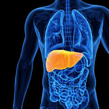 Drug-induced hepatitis (DIH) is one of the major complications among the treatment of patients with tuberculosis (TB); it might even be fatal. This study tries to address the recurrence of DIH with 2 anti-TB regimens.
In the retrospective study from 2007 to 2010, 135 TB patients with DIH who were older than 16 years were entered to study. The patients with DIH were randomly treated with a regimen, including isoniazid, rifampin, and ethambutol, plus either ofloxacin or pyrazinamide. The patients were reviewed for occurrence of recurrent DIH. Cure and completed treatment were considered as acceptable treatment outcomes, whereas default of treatment, treatment failure, and death were considered to be unacceptable outcomes. Therefore, 135 subjects with DIH were reviewed, and 23 patients (17%) experienced recurrence of hepatitis (19 cases in the ofloxacin group and 4 cases in the pyrazinamide group). There is no significant difference in recurrence of hepatitis between these 2 groups (P = 0.803). An acceptable outcome was observed in 95 patients (70.4%), and an unacceptable outcome was seen in 14 cases (10.3%). There was no significant difference in outcomes between these 2 regimens (P = 0.400, odds ratio = 1.62, 95% confidence interval, 0.524-4.98).
The results of our study suggest that ofloxacin-based anti-TB regimen does not decrease the risk of recurrent DIH. Therefore, adding ofloxacin in the case of DIH is not recommended.
Interstitial lung disease related to smoking: imaging considerations
 Purpose of review: To discuss the imaging of interstitial lung disease believed to be caused by smoking.
Recent findings: It is increasingly clear that smoking is associated with a variety of patterns of interstitial lung disease. The radiologic features of interstitial lung disease caused by smoking cigarettes are variable and may be nonspecific.
Summary: It is now accepted that cigarette smoking can cause lung diseases other than lung cancer, chronic bronchitis and emphysema. Indeed, the hypothesis that tobacco smoke can cause interstitial lung disease – and, specifically, pulmonary fibrosis – dates back to the 1960s. The list of interstitial lung disease, in which smoking is believed to have an etiologic role, includes Langerhans’ cell histiocytosis, respiratory bronchiolitis/respiratory bronchiolitis-interstitial lung disease and desquamative interstitial pneumonia. More recently, there is emerging evidence which suggests that smoking may be associated with other patterns of pulmonary fibrosis (e.g. nonspecific interstitial pneumonia and smoking-related interstitial fibrosis). In the present review we discuss the imaging of the interstitial lung disease known to be caused by smoking; the typical appearances and some of the diagnostic difficulties are discussed.
Purpose of review: To discuss the imaging of interstitial lung disease believed to be caused by smoking.
Recent findings: It is increasingly clear that smoking is associated with a variety of patterns of interstitial lung disease. The radiologic features of interstitial lung disease caused by smoking cigarettes are variable and may be nonspecific.
Summary: It is now accepted that cigarette smoking can cause lung diseases other than lung cancer, chronic bronchitis and emphysema. Indeed, the hypothesis that tobacco smoke can cause interstitial lung disease – and, specifically, pulmonary fibrosis – dates back to the 1960s. The list of interstitial lung disease, in which smoking is believed to have an etiologic role, includes Langerhans’ cell histiocytosis, respiratory bronchiolitis/respiratory bronchiolitis-interstitial lung disease and desquamative interstitial pneumonia. More recently, there is emerging evidence which suggests that smoking may be associated with other patterns of pulmonary fibrosis (e.g. nonspecific interstitial pneumonia and smoking-related interstitial fibrosis). In the present review we discuss the imaging of the interstitial lung disease known to be caused by smoking; the typical appearances and some of the diagnostic difficulties are discussed.Video-assisted thoracoscopic surgery for retained hemothorax in blunt chest trauma
 Purpose of review: In the last decade, video-assisted thoracoscopic surgery (VATS) has become a popular method in diagnosis and treatment of acute chest injuries. Except for patients with unstable vital signs who require larger surgical incisions to check bleeding, this endoscopic surgery could be employed in the majority of thoracic injury patients with stable vital signs.
Recent findings: In the past, VATS was used to evacuate traumatic-retained hemothorax. Recent study has revealed further that lung repair during VATS could decrease complications after trauma. Management of fractured ribs could also be assisted by VATS. Early VATS intervention within 7 days after injury can decrease the rate of posttraumatic infection and length of hospital stay. In studies of the pathophysiology of animal models, N-acetylcysteine and methylene blue were used in animals with blunt chest trauma and found to improve clinical outcomes.
Summary: Retained hemothorax derived from blunt chest trauma should be managed carefully and rapidly. Early VATS intervention is a well tolerated and reliable procedure that can be applied to manage this complication cost effectively.
Purpose of review: In the last decade, video-assisted thoracoscopic surgery (VATS) has become a popular method in diagnosis and treatment of acute chest injuries. Except for patients with unstable vital signs who require larger surgical incisions to check bleeding, this endoscopic surgery could be employed in the majority of thoracic injury patients with stable vital signs.
Recent findings: In the past, VATS was used to evacuate traumatic-retained hemothorax. Recent study has revealed further that lung repair during VATS could decrease complications after trauma. Management of fractured ribs could also be assisted by VATS. Early VATS intervention within 7 days after injury can decrease the rate of posttraumatic infection and length of hospital stay. In studies of the pathophysiology of animal models, N-acetylcysteine and methylene blue were used in animals with blunt chest trauma and found to improve clinical outcomes.
Summary: Retained hemothorax derived from blunt chest trauma should be managed carefully and rapidly. Early VATS intervention is a well tolerated and reliable procedure that can be applied to manage this complication cost effectively.